
The foot and ankle are intricate and essential parts of the human body, working together as complex joints. They are crucial for movement, balance, stability, and supporting the entire body’s weight. Understanding the anatomy of your feet and ankles is the first step in appreciating their function and knowing when to seek expert care if you experience pain or problems. If you’re experiencing discomfort, searching for a “Foot Ankle Doctor Near Me” is a crucial step towards relief.
 3D anatomy illustration of the human foot and ankle bones, highlighting the complex structure. For foot and ankle pain, consult a foot ankle doctor near me.
The foot and ankle are composed of a remarkable assembly of 26 bones, 33 joints, and a network of muscles, tendons, and ligaments, all working in harmony. Let’s delve into the specifics of this anatomy.
Bones of the Ankle
The ankle joint serves as the critical connection point between the leg and the foot. This joint is formed by three bones:
- Tibia: Also known as the shinbone, it’s the larger bone of the lower leg.
- Fibula: Referred to as the calf bone, it’s the smaller bone of the lower leg.
- Talus: The ankle bone itself, which articulates with the tibia and fibula, enabling the foot’s up and down motion, essential for walking, running, and jumping.
Key bony landmarks that form the ankle joint are the malleoli, bony bumps located at the ends of the tibia and fibula:
- Medial Malleolus: This is the bump on the inside of your ankle, formed by the tibia.
- Posterior Malleolus: Located at the back of the ankle, this is also a part of the tibia.
- Lateral Malleolus: Found on the outer side of the ankle, this prominent bump is formed by the fibula.
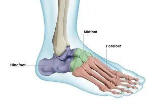
Bones of the Foot
While the foot functions as a unified structure, it can be anatomically divided into three sections: the hindfoot, midfoot, and forefoot.
-
Hindfoot: Forming the foundation of the ankle and heel, the hindfoot consists of two primary bones:
- Talus: Shared with the ankle joint.
- Calcaneus: Also known as the heel bone, it is the largest bone in the foot and bears a significant amount of weight.
-
Midfoot: This section acts as a bridge connecting the hindfoot and forefoot. It is comprised of five bones:
- Navicular Bone: Situated in front of the heel bone.
- Cuboid Bone: Located on the outer side of the midfoot.
- Three Cuneiform Bones: Medial, intermediate, and lateral cuneiform bones arranged in front of the navicular bone, contributing to the arch of the foot.
-
Forefoot: The forefoot includes the metatarsals and phalanges, forming the toes and the ball of the foot.
- Metatarsals: Five metatarsal bones extend from the midfoot to the base of the toes, forming the arch of the foot. This arch is vital for shock absorption during activities like walking and running.
- Phalanges: These are the bones of the toes. Each toe has three phalanges (proximal, middle, and distal), except for the big toe (hallux), which has only two (proximal and distal).
- Sesamoid Bones: The big toe also contains two small, round sesamoid bones located in the ball of the foot beneath the joint of the big toe. These bones assist in the upward and downward movement of the big toe and reduce pressure on the tendons.
Ankle and Foot Joints
The foot and ankle contain a total of 33 joints, allowing for a wide range of motion and flexibility. These joints can be categorized into:
- Hinge Joints: The primary ankle joint is a hinge joint, primarily allowing for dorsiflexion (bending the foot upwards) and plantarflexion (pointing the foot downwards).
- Gliding Joints: Found in the hindfoot, particularly between the talus and calcaneus, these joints enable gliding and side-to-side movements, contributing to foot flexibility on uneven surfaces.
- Condyloid Joints: Located in the forefoot at the metatarsophalangeal joints (where toes meet the metatarsals) and interphalangeal joints (within the toes), these joints allow for flexion, extension, abduction (moving away from the midline of the body), and adduction (moving towards the midline).
These numerous joints work together to provide stability, support body weight, and enable adaptation to varied terrains while walking, running, or performing other activities.
Soft Tissues of the Ankle and Foot
Beyond bones and joints, soft tissues are crucial for the function and stability of the foot and ankle. These include cartilage, ligaments, muscles, tendons, and bursae.
-
Articular Cartilage: The ends of all bones within the ankle and foot joints are covered with articular cartilage. This smooth, resilient tissue acts as a cushion, reducing friction between bones during movement and absorbing shock. Synovial fluid further lubricates these joints, promoting smooth, pain-free motion.
-
Ligaments: These strong, fibrous tissues connect bones to bones, providing stability and limiting excessive joint movement. Key ligaments include:
- Plantar Fascia: The largest ligament in the foot, it runs along the bottom of the foot from the heel to the forefoot, supporting the arch and acting as a shock absorber.
- Lateral Ligaments: Located on the outside of the ankle, these ligaments (anterior talofibular, calcaneofibular, and posterior talofibular ligaments) prevent excessive inversion (rolling outwards) of the ankle.
- Medial Ligaments (Deltoid Ligaments): Situated on the inside of the ankle, these ligaments are stronger and prevent excessive eversion (rolling inwards).
-
Muscles: Twenty muscles within the foot and ankle are responsible for movement, support, and maintaining the foot’s arches. Key muscle groups include:
- Anterior Tibial Muscle: Facilitates dorsiflexion of the foot.
- Posterior Tibial Muscle: Supports the arch of the foot and aids in plantarflexion and inversion.
- Peroneal Muscles (Peroneus Longus and Brevis): Control movement on the outside of the ankle, responsible for eversion and plantarflexion.
- Extensor Muscles (Extensor Digitorum Longus and Hallucis Longus): Raise the toes and foot during swing phase of walking.
- Flexor Muscles (Flexor Digitorum Longus and Hallucis Longus): Stabilize the toes against the ground and assist in plantarflexion.
- Intrinsic Foot Muscles: Smaller muscles within the foot that fine-tune toe movements and support the arches.
-
Tendons: Tendons are tough, fibrous cords that connect muscles to bones, transmitting the force of muscle contraction to create movement.
- Achilles Tendon: The strongest and largest tendon in the body, connecting the calf muscles to the heel bone, essential for powerful plantarflexion (pushing off when walking or running).
- Peroneal Tendons: Attach peroneal muscles to foot bones.
- Tibialis Anterior and Posterior Tendons: Attach tibialis anterior and posterior muscles to foot bones.
-
Bursae: These small, fluid-filled sacs are located around joints, tendons, and bones, reducing friction and allowing smooth movement between these structures. They contain synovial fluid similar to that in joints, further minimizing friction.
Understanding the complex anatomy of the foot and ankle highlights their incredible design for mobility and support. If you experience pain, injury, or discomfort in your feet or ankles, seeking professional evaluation is essential. Search for a “foot ankle doctor near me” to find qualified specialists who can diagnose and treat your specific needs, ensuring you maintain healthy and functional feet and ankles for an active life.