
Can an eye doctor detect a tumor behind your eye? Yes, an eye doctor, or ophthalmologist, can often detect a tumor behind the eye during a comprehensive eye exam. At thebootdoctor.net, we emphasize the importance of regular eye check-ups for maintaining overall health and identifying potential issues early. These examinations not only assess your vision but also allow the doctor to examine the structures of the eye and surrounding areas for any abnormalities, providing timely diagnosis and care. Early detection and intervention can significantly improve outcomes for various eye and vision concerns.
Discover more eye care insights on our platform, including details on neuro-ophthalmic exams, visual field testing, and optic nerve health.
1. What Types of Tumors Can an Eye Exam Detect?
An eye exam can detect various types of tumors, both within the eye and in the surrounding areas. Eye doctors, or ophthalmologists, are trained to identify abnormalities during routine and specialized examinations. Here’s a detailed look at the types of tumors that can be detected:
- Retinoblastoma: This is a rare malignant tumor of the retina, most commonly affecting young children. According to the American Academy of Ophthalmology, early detection through a comprehensive eye exam can significantly improve the chances of successful treatment.
- Choroidal Melanoma: This is the most common primary intraocular malignancy in adults. It arises from the pigment cells (melanocytes) in the choroid, the vascular layer beneath the retina. Regular eye exams can help in early detection and management.
- Optic Nerve Tumors: Tumors affecting the optic nerve, such as optic nerve gliomas or meningiomas, can cause vision changes and abnormalities detectable during an eye exam.
- Orbital Tumors: These tumors occur in the tissues surrounding the eye within the bony socket (orbit). They can be benign or malignant and may affect vision, eye movement, and the appearance of the eye.
- Pituitary Tumors: Although located in the brain, pituitary tumors can affect vision due to their proximity to the optic nerve and chiasm. An eye exam, particularly a visual field test, can help detect these effects.
Understanding the types of tumors that can be detected through an eye exam highlights the importance of regular eye check-ups. Thebootdoctor.net offers resources and information to help you stay informed and proactive about your eye health.
2. How Does an Eye Doctor Check for Tumors Behind the Eye?
Eye doctors employ several specialized techniques to check for tumors behind the eye during a comprehensive eye exam. These methods allow them to visualize the structures of the eye and detect any abnormalities that may indicate the presence of a tumor. Here are some key procedures:
- Ophthalmoscopy: This involves using an ophthalmoscope to examine the retina, optic nerve, and blood vessels at the back of the eye. Dilating eye drops are often used to widen the pupil, providing a better view.
- Slit-Lamp Examination: A slit lamp is a microscope with a bright light used to examine the front structures of the eye, including the eyelids, cornea, iris, and lens. It can also be used with special lenses to view the retina and optic nerve.
- Tonometry: This test measures the pressure inside the eye (intraocular pressure) and is primarily used to screen for glaucoma, but it can also provide information about the overall health of the eye.
- Visual Field Testing: This test assesses the extent of your peripheral vision. It can help detect blind spots or visual field defects caused by tumors pressing on the optic nerve or affecting the visual pathways in the brain.
- Optical Coherence Tomography (OCT): OCT is an imaging technique that uses light waves to take cross-sectional pictures of your retina. It can reveal abnormalities in the retinal layers and optic nerve, aiding in the detection of tumors and other conditions.
- Fundus Photography: This involves taking detailed photographs of the back of the eye, including the retina, optic disc, and blood vessels. These images can be used to document the appearance of the eye and monitor changes over time.
- Fluorescein Angiography: This is a diagnostic procedure that uses a fluorescent dye to highlight blood vessels in the retina and choroid. It can help detect abnormalities in blood flow caused by tumors or other vascular conditions.
Regular eye exams at facilities like thebootdoctor.net include these tests to ensure thorough monitoring of your eye health.
3. What are the Symptoms That Might Indicate a Tumor Behind the Eye?
Several symptoms might indicate the presence of a tumor behind the eye. Recognizing these signs early is crucial for timely diagnosis and treatment. Here are some key symptoms to watch out for:
- Vision Changes:
- Blurred Vision: Difficulty seeing clearly, as if the image is out of focus.
- Double Vision (Diplopia): Seeing two images of a single object.
- Loss of Peripheral Vision: Difficulty seeing objects to the side, resulting in a narrowed field of view.
- Decreased Visual Acuity: A gradual or sudden reduction in the sharpness of vision.
- Eye Pain or Discomfort:
- Persistent Eye Pain: A constant ache or soreness in or around the eye.
- Pain with Eye Movement: Discomfort or pain that worsens when moving the eyes.
- Proptosis (Bulging of the Eye):
- Visible Protrusion: The eye appearing to bulge or protrude from the eye socket.
- Changes in Pupil Size or Shape:
- Unequal Pupil Size (Anisocoria): One pupil being noticeably larger or smaller than the other.
- Pupil Shape Irregularities: The pupil appearing misshapen or distorted.
- Eye Movement Problems:
- Difficulty Moving the Eye: Restricted or impaired eye movement in one or more directions.
- Misalignment of the Eyes (Strabismus): The eyes not pointing in the same direction.
- Headaches:
- Persistent Headaches: Frequent or constant headaches, especially if they are new or different from previous headaches.
- Headaches Accompanied by Vision Changes: Headaches that occur with blurred vision, double vision, or other visual disturbances.
- Swelling or Mass Around the Eye:
- Visible Lump: A noticeable swelling or mass in the tissues around the eye.
- Other Neurological Symptoms:
- Nausea and Vomiting: Feeling sick to the stomach, especially if accompanied by headaches or vision changes.
- Dizziness: Feeling lightheaded or unsteady.
- Seizures: Uncontrolled electrical disturbances in the brain.
According to the Mayo Clinic, individuals experiencing any of these symptoms should seek prompt medical attention for a thorough evaluation. Regular check-ups at places like thebootdoctor.net can also aid in early detection and management.
4. Can Routine Eye Exams Detect Early Signs of a Brain Tumor?
Yes, routine eye exams can often detect early signs of a brain tumor. While these exams are primarily focused on assessing vision and eye health, they can also reveal abnormalities that may indicate the presence of a brain tumor, particularly those affecting the optic nerve or surrounding areas.
- Visual Field Defects: Brain tumors that press on the optic nerve or visual pathways in the brain can cause characteristic patterns of visual field loss.
- Optic Disc Swelling (Papilledema): Increased pressure inside the skull (intracranial pressure) due to a brain tumor can cause swelling of the optic disc, the area where the optic nerve enters the eye.
- Abnormal Eye Movements: Tumors affecting the cranial nerves that control eye movement can cause misalignment of the eyes (strabismus), double vision (diplopia), or other abnormalities in eye movement.
The American Academy of Ophthalmology emphasizes that early detection of these signs during a routine eye exam can lead to earlier diagnosis and treatment of brain tumors, potentially improving outcomes. Regular eye check-ups at thebootdoctor.net can help in identifying these early signs.
5. What Specific Tests are Conducted During an Eye Exam to Look for Tumors?
During an eye exam to check for tumors, eye doctors conduct several specific tests to evaluate the structures of the eye and detect any abnormalities. These tests help in visualizing the retina, optic nerve, and surrounding tissues, allowing for early detection of potential tumors. Here are the key tests performed:
- Visual Acuity Test: This measures the sharpness and clarity of your vision at various distances. It involves reading letters or symbols on a standardized eye chart (Snellen chart).
- Refraction: This test determines your refractive error (nearsightedness, farsightedness, astigmatism) and helps in prescribing the appropriate corrective lenses (glasses or contact lenses).
- Visual Field Test: This assesses the extent of your peripheral vision and detects any blind spots or visual field defects.
- Pupil Examination: This evaluates the size, shape, and reactivity of your pupils to light. Abnormalities in pupil size or reactivity can indicate neurological issues.
- Eye Movement Assessment: This assesses the alignment and movement of your eyes in all directions. Abnormalities in eye movement can suggest problems with the cranial nerves or muscles controlling eye movement.
- Slit-Lamp Examination: This involves using a slit lamp (a microscope with a bright light) to examine the front structures of the eye, including the eyelids, cornea, iris, and lens.
- Ophthalmoscopy (Fundoscopy): This uses an ophthalmoscope to examine the back of the eye, including the retina, optic disc, and blood vessels.
- Tonometry: This measures the pressure inside the eye (intraocular pressure).
- Optical Coherence Tomography (OCT): OCT is an imaging technique that uses light waves to take cross-sectional pictures of your retina.
- Fundus Photography: This involves taking detailed photographs of the back of the eye.
Thebootdoctor.net recommends regular eye exams to include these tests, ensuring comprehensive monitoring of your eye health and early detection of any potential issues.
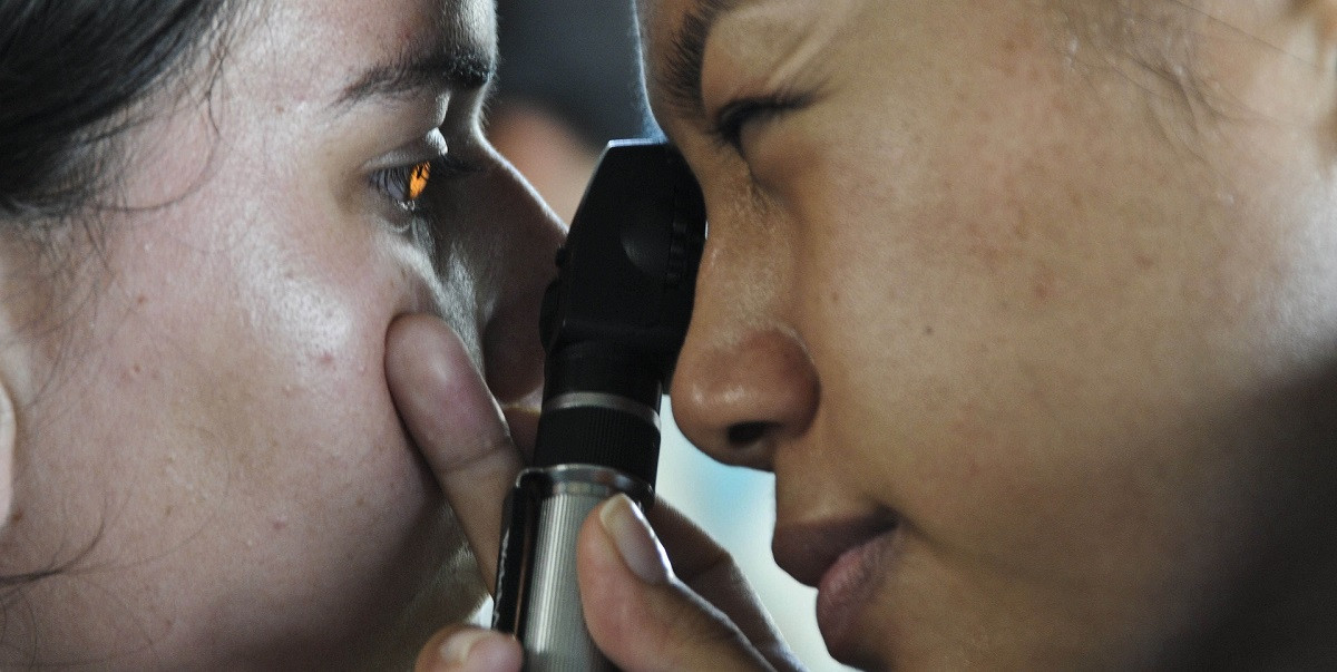 Optometrist using ophthalmoscope to check for tumors
6. How Often Should You Get an Eye Exam to Ensure Early Detection?
The frequency of eye exams for early tumor detection varies depending on age, risk factors, and overall health. Regular eye check-ups are essential for maintaining eye health and detecting potential problems early. Here are general guidelines for eye exam frequency:
- Children:
- The American Academy of Ophthalmology recommends that children have their first comprehensive eye exam at 6 months of age, another at age 3, and again before starting school (around age 5 or 6).
- School-age children should have an eye exam every 1-2 years, or more frequently if recommended by their eye doctor.
- Adults:
- Ages 20-39: Adults with no known risk factors or vision problems should have an eye exam at least once every 5-10 years.
- Ages 40-54: Adults in this age group should have an eye exam every 2-4 years.
- Ages 55-64: Eye exams should be conducted every 1-3 years.
- Ages 65 and Older: Annual eye exams are recommended.
- Individuals with Risk Factors:
- More frequent eye exams are recommended for individuals with certain risk factors, such as:
- Family History of Eye Disease: Glaucoma, macular degeneration, or other hereditary eye conditions.
- Diabetes: Diabetic retinopathy can lead to vision loss if not detected and managed early.
- High Blood Pressure: Hypertension can damage blood vessels in the eye, leading to retinopathy.
- Previous Eye Injury or Surgery: History of trauma or surgical procedures to the eye.
- Use of Certain Medications: Some medications can have side effects that affect the eyes.
- Wearing Contact Lenses: Regular check-ups are necessary to monitor corneal health and prevent complications.
- More frequent eye exams are recommended for individuals with certain risk factors, such as:
Adhering to these guidelines and consulting with eye care professionals like those at thebootdoctor.net ensures timely detection and management of eye conditions, including tumors.
7. What Happens if an Eye Doctor Suspects a Tumor During an Exam?
If an eye doctor suspects a tumor during an eye exam, they will typically take several steps to confirm the diagnosis and determine the appropriate course of action. Here’s a detailed overview of the process:
- Further Diagnostic Testing:
- Imaging Studies:
- Magnetic Resonance Imaging (MRI): MRI provides detailed images of the brain and surrounding structures.
- Computed Tomography (CT) Scan: CT scans use X-rays to create cross-sectional images of the brain and skull.
- Ultrasound: Ultrasound uses sound waves to create images of the eye and orbit.
- Visual Field Testing:
- Repeat or more detailed visual field testing may be performed to assess the extent of any visual field loss.
- Optical Coherence Tomography (OCT):
- OCT scans provide high-resolution images of the retina and optic nerve, helping to detect subtle abnormalities.
- Fluorescein Angiography:
- This test involves injecting a fluorescent dye into a vein and taking photographs of the blood vessels in the retina.
- Imaging Studies:
- Referral to a Specialist:
- Neuro-Ophthalmologist:
- This is an ophthalmologist with specialized training in neurological conditions affecting the eyes and vision.
- Ophthalmologist with Expertise in Ocular Oncology:
- This is an ophthalmologist who specializes in the diagnosis and treatment of eye tumors.
- Neurosurgeon:
- A neurosurgeon may be consulted if a brain tumor is suspected, particularly if it is affecting the optic nerve or visual pathways.
- Neuro-Ophthalmologist:
- Biopsy (If Necessary):
- In some cases, a biopsy may be necessary to confirm the diagnosis and determine the type of tumor.
- Treatment Planning:
- Once a diagnosis is confirmed, a treatment plan will be developed based on the type, size, and location of the tumor, as well as the patient’s overall health.
- Surgical Removal: Surgical removal of the tumor may be possible, depending on its location and size.
- Radiation Therapy: Radiation therapy uses high-energy rays to kill tumor cells.
- Chemotherapy: Chemotherapy involves using drugs to kill tumor cells.
- Targeted Therapy: Targeted therapy uses drugs that specifically target cancer cells without harming normal cells.
- Observation: In some cases, small, slow-growing tumors may be monitored over time without immediate intervention.
Thebootdoctor.net emphasizes the importance of following the eye doctor’s recommendations and seeking prompt medical attention if a tumor is suspected.
8. What are the Risk Factors for Developing Tumors Behind the Eye?
Several risk factors can increase the likelihood of developing tumors behind the eye. Understanding these factors can help individuals and healthcare providers monitor and manage potential risks. Here are some of the key risk factors:
- Genetic Predisposition:
- Hereditary Conditions: Certain genetic conditions can increase the risk of developing tumors.
- Neurofibromatosis Type 1 (NF1): This genetic disorder causes tumors to grow along nerves throughout the body, including the optic nerve.
- Retinoblastoma: A rare childhood cancer of the retina is often caused by a genetic mutation.
- Age:
- Children: Retinoblastoma is most common in young children.
- Adults: Other types of tumors, such as optic nerve meningiomas, are more common in adults.
- Gender:
- Women: Meningiomas, which can affect the optic nerve, are more common in women than in men.
- Race and Ethnicity:
- Certain Populations: Some studies suggest that certain racial and ethnic groups may have a higher risk of specific types of tumors.
- Medical History:
- Previous Cancer: Individuals with a history of cancer, particularly those who have received radiation therapy to the head or neck, may have an increased risk of developing tumors.
- Environmental Factors:
- Radiation Exposure: Exposure to ionizing radiation, such as from radiation therapy, can increase the risk of developing tumors.
- Chemical Exposure: Exposure to certain chemicals may also play a role in increasing tumor risk, although more research is needed in this area.
- Immune System Disorders:
- Weakened Immune System: Individuals with weakened immune systems, such as those with HIV/AIDS or those taking immunosuppressant drugs, may have an increased risk of developing certain types of tumors.
- Other Medical Conditions:
- Tuberous Sclerosis: This genetic disorder can cause tumors to grow in various organs, including the eyes and brain.
- Von Hippel-Lindau (VHL) Disease: This genetic disorder increases the risk of developing tumors and cysts in various parts of the body, including the eyes and brain.
According to the National Institutes of Health (NIH), being aware of these risk factors and discussing them with your healthcare provider can help in early detection and management. Thebootdoctor.net provides resources for staying informed about eye health and potential risks.
9. Can Tumors Behind the Eye Cause Other Health Problems?
Yes, tumors behind the eye can cause a range of other health problems, depending on their size, location, and growth rate. These problems can affect vision, neurological function, and overall quality of life. Here are some of the potential health issues associated with tumors behind the eye:
- Vision Loss:
- Gradual Vision Loss: Tumors pressing on the optic nerve can cause a slow, progressive decline in vision.
- Sudden Vision Loss: In some cases, tumors can cause sudden vision loss due to compression or disruption of blood flow to the optic nerve.
- Peripheral Vision Loss: Tumors can affect the peripheral visual field, leading to tunnel vision.
- Double Vision (Diplopia): Tumors affecting the cranial nerves that control eye movement can cause double vision.
- Eye Movement Problems:
- Strabismus (Misalignment of the Eyes): Tumors can cause the eyes to become misaligned, leading to double vision and other visual disturbances.
- Nystagmus (Involuntary Eye Movements): Tumors affecting the brainstem or cerebellum can cause involuntary, rhythmic eye movements.
- Headaches:
- Persistent Headaches: Tumors can cause persistent headaches, which may be worse in the morning or accompanied by nausea and vomiting.
- Neurological Symptoms:
- Seizures: Tumors in the brain can cause seizures, which are characterized by uncontrolled electrical activity in the brain.
- Weakness or Paralysis: Tumors can affect motor function, leading to weakness or paralysis on one side of the body.
- Cognitive Changes: Tumors can cause changes in cognitive function, such as memory loss, difficulty concentrating, or changes in personality.
- Hormonal Imbalances:
- Pituitary Tumors: Tumors in the pituitary gland can disrupt the production of hormones, leading to hormonal imbalances that can affect various bodily functions.
- Increased Intracranial Pressure:
- Hydrocephalus: Tumors can block the flow of cerebrospinal fluid, leading to a buildup of fluid in the brain (hydrocephalus) and increased intracranial pressure.
- Papilledema: Increased intracranial pressure can cause swelling of the optic disc, which can be detected during an eye exam.
- Other Systemic Effects:
- Fatigue: Tumors can cause fatigue and a general feeling of unwellness.
- Weight Loss: Tumors can lead to unintentional weight loss.
- Nausea and Vomiting: Tumors can cause nausea and vomiting, particularly if they are located in the brainstem or cerebellum.
According to the American Brain Tumor Association, early detection and treatment of tumors behind the eye can help prevent or minimize these complications. Thebootdoctor.net emphasizes the importance of regular check-ups.
10. What Advances Have Been Made in Detecting and Treating These Tumors?
Significant advances have been made in recent years in both the detection and treatment of tumors behind the eye. These advancements have led to earlier diagnosis, more effective treatments, and improved outcomes for patients. Here are some of the key developments:
- Advanced Imaging Techniques:
- High-Resolution MRI: Improved MRI technology provides more detailed images of the brain and surrounding structures.
- Optical Coherence Tomography (OCT): OCT provides high-resolution images of the retina and optic nerve, allowing for early detection of subtle abnormalities.
- PET Scans: Positron emission tomography (PET) scans can help differentiate between benign and malignant tumors.
- Genetic Testing:
- Next-Generation Sequencing (NGS): NGS allows for rapid and comprehensive genetic testing of tumor samples, providing valuable information about the tumor’s molecular characteristics.
- Targeted Therapies:
- Monoclonal Antibodies: These drugs target specific proteins on tumor cells, disrupting their growth and spread.
- Small Molecule Inhibitors: These drugs inhibit specific signaling pathways involved in tumor growth and survival.
- Immunotherapy:
- Checkpoint Inhibitors: These drugs block proteins that prevent the immune system from attacking tumor cells.
- CAR-T Cell Therapy: This involves modifying a patient’s T cells to recognize and attack tumor cells.
- Surgical Techniques:
- Minimally Invasive Surgery: Minimally invasive surgical techniques allow for the removal of tumors through small incisions.
- Image-Guided Surgery: This technique uses real-time imaging to guide the surgeon during tumor removal, improving precision and minimizing damage to surrounding tissues.
- Radiation Therapy:
- Stereotactic Radiosurgery (SRS): SRS delivers high doses of radiation to a small, targeted area.
- Proton Therapy: Proton therapy uses protons instead of X-rays to deliver radiation, reducing the risk of damage to surrounding tissues.
- Clinical Trials:
- Ongoing clinical trials are evaluating new and innovative approaches to the detection and treatment of tumors behind the eye.
Thebootdoctor.net stays updated on these advancements, providing our readers with the latest information on eye health and tumor detection and treatment.
FAQ: Eye Tumors and Detection
- Can an eye doctor detect a brain tumor?
- Yes, an eye doctor can often detect signs of a brain tumor during a comprehensive eye exam by observing changes in the optic nerve or visual field.
- What are the early symptoms of a tumor behind the eye?
- Early symptoms may include blurred vision, double vision, loss of peripheral vision, eye pain, and headaches.
- How often should I get an eye exam to check for tumors?
- Adults should get an eye exam every 1-3 years, depending on age and risk factors; children should have exams at 6 months, 3 years, and before starting school.
- What tests are performed during an eye exam to look for tumors?
- Tests include visual acuity, visual field testing, pupil examination, ophthalmoscopy, and optical coherence tomography (OCT).
- What happens if my eye doctor suspects a tumor?
- The eye doctor will refer you to a specialist for further diagnostic testing, such as MRI or CT scans, to confirm the diagnosis.
- Are there risk factors for developing tumors behind the eye?
- Risk factors include genetic predispositions, age, gender, race, medical history, and exposure to radiation.
- Can tumors behind the eye cause other health problems?
- Yes, they can cause vision loss, eye movement problems, headaches, neurological symptoms, and hormonal imbalances.
- What is the role of imaging tests like MRI and CT scans?
- MRI and CT scans provide detailed images of the brain and surrounding structures, helping to confirm the presence and location of tumors.
- What are the treatment options for tumors behind the eye?
- Treatment options include surgical removal, radiation therapy, chemotherapy, targeted therapy, and immunotherapy.
- How can I stay informed about eye health and tumor detection?
- Regular eye exams, consulting with eye care professionals, and staying informed through resources like thebootdoctor.net can help.
At thebootdoctor.net, we are dedicated to providing comprehensive and reliable information on eye health. If you have any concerns or questions, please don’t hesitate to contact our team or schedule an appointment with an eye care professional.
Address: 6565 Fannin St, Houston, TX 77030, United States
Phone: +1 (713) 791-1414
Website: thebootdoctor.net