
Bladder cancer is a significant health concern affecting many individuals annually. An early bladder cancer diagnosis is paramount for improving treatment outcomes and survival rates. Recognizing the symptoms of bladder cancer, such as blood in your urine, increased urinary frequency, or pain when urinating, is the first step toward timely detection and effective intervention. If you experience any of these symptoms, it’s crucial to consult a healthcare professional, specifically a Bladder Doctor, without delay.
When bladder cancer is suspected, a series of diagnostic evaluations will be performed by your bladder doctor to confirm the diagnosis. These may include urine tests, cystoscopy, and various imaging scans. These tests are essential not only for diagnosis but also for determining the cancer’s stage and extent, which is critical for formulating an appropriate treatment plan. The earlier bladder cancer is diagnosed by a skilled bladder doctor, the greater the likelihood of successful treatment and recovery. Below, we delve deeper into the diagnostic steps involved in bladder cancer detection and highlight when seeking the expertise of a bladder doctor is essential.
Your bladder doctor will begin by taking a detailed medical history. This will include questions about your symptoms, existing risk factors for bladder cancer, and any family history of the disease. This initial consultation is a crucial step in determining the need for further diagnostic testing.
Essential Tests Your Bladder Doctor Uses for Bladder Cancer Diagnosis
Several tests are available to bladder doctors to effectively diagnose bladder cancer. These tests range from simple urine analysis to more complex imaging and invasive procedures, each playing a vital role in accurate diagnosis.
Urinalysis: The First Step
One of the most frequently reported symptoms of bladder cancer and a key indicator for your bladder doctor is blood in the urine, also known as hematuria. While hematuria can be caused by various conditions, it warrants immediate investigation by a bladder doctor to rule out serious conditions like bladder cancer.
A specific urine test, known as urine cytology, is often performed by bladder doctors. This test examines a urine sample under a microscope to detect any abnormal cells. In some cases, if unusual cells are identified, more advanced analysis such as molecular analysis may be conducted to identify tumor cells in the urine with greater precision.
Cystoscopy: Direct Visualization by Your Bladder Doctor
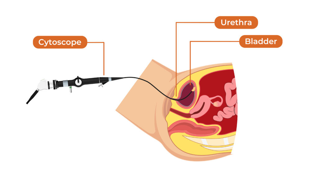
Cystoscopy is a cornerstone procedure in the diagnosis of bladder cancer and is expertly performed by bladder doctors. This diagnostic procedure involves inserting a cystoscope – a thin, flexible tube equipped with a camera – through the urethra and into the bladder. This allows the bladder doctor to directly visualize the bladder lining and identify any abnormal growths, lesions, or irregularities. If any suspicious areas are observed during the cystoscopy, the bladder doctor can perform a biopsy to obtain tissue samples for further examination and definitive diagnosis. While cystoscopy is highly effective in detecting bladder abnormalities, a biopsy is often necessary to confirm a cancer diagnosis. A bladder doctor’s expertise in performing and interpreting cystoscopy is crucial for early and accurate detection.
Biopsy: Confirming the Diagnosis
A biopsy, typically performed during a cystoscopy by your bladder doctor, is essential for confirming a diagnosis of bladder cancer. This procedure involves removing a small tissue sample from any suspicious growth identified in the bladder. If a tumor or other abnormality is detected during cystoscopy, the bladder doctor, often a urologist specializing as a bladder doctor, will schedule a cystoscopy under anesthesia specifically to perform a bladder biopsy. During this biopsy, the bladder doctor will carefully remove tissue samples and send them to a pathology lab for detailed analysis by a pathologist. To ensure comprehensive assessment, the bladder doctor may take multiple biopsies from different areas of the growth.
Following the biopsy, the bladder doctor may perform a transurethral resection of the bladder tumor, or TURBT. TURBT is a procedure where the bladder doctor uses specialized instruments inserted through the urethra to remove as much of the tumor as possible. The removed tumor tissue is then sent for pathological examination to determine the cancer stage and grade.
In addition to bladder-specific tests, your bladder doctor may order further tests to evaluate other parts of your urinary tract to gain a complete understanding of your condition.
CT Urogram: Imaging the Upper Urinary Tract
A CT urogram is a specialized radiological test that bladder doctors use to investigate the causes of blood in the urine or other urinary symptoms. This scan uses intravenous (IV) contrast dye to enhance the visualization of internal structures in X-ray imaging. A CT urogram provides detailed images of the upper urinary tract, including the kidneys and ureters.
This test is particularly effective for bladder doctors in detecting tumors in the kidney, renal pelvis, and ureter, as well as identifying other urologic abnormalities. It can also reveal kidney stones and hydronephrosis , which is kidney swelling often caused by urinary blockage. Furthermore, the CT urogram images the entire abdomen and pelvis, allowing the bladder doctor and radiologist to identify abnormalities in these regions, including signs of cancer spread to lymph nodes or other organs in patients with known cancer.
Before a CT urogram with contrast can be performed, your healthcare provider, including your bladder doctor, will order blood work to assess your kidney function. If contrast dye cannot be safely administered, the bladder doctor may opt for a CT scan without contrast or consider alternative imaging methods. In some cases, a cystoscopy with retrograde pyelograms might be recommended. During this procedure, the bladder doctor injects dye directly into the ureters during cystoscopy while taking X-rays, similar to a CT urogram, to visualize the ureters and renal pelvis for abnormalities.
It’s important to note that while CT urograms and other imaging tests can detect some bladder tumors, they may not identify all. Therefore, a bladder doctor will often recommend a cystoscopy to directly examine the lower urinary tract (bladder and urethra) for any source of blood in the urine or to investigate other urologic symptoms comprehensively.
MR Urogram: An Alternative Imaging Method
Another imaging option available to bladder doctors is an MRI of the abdomen and pelvis, or MR Urogram. This test is also proficient in detecting tumors within the kidney and ureters and identifying potential cancer spread. MR Urograms are particularly useful in situations where radiation exposure should be minimized or for patients with contrast dye allergies or compromised kidney function. However, it is less effective at detecting kidney stones compared to CT urograms and, like CT urograms, may miss some bladder tumors, necessitating a cystoscopy for complete evaluation by a bladder doctor.
Renal Ultrasound: A Non-invasive Approach
Renal ultrasound is the least invasive imaging technique for evaluating the kidneys and can be utilized by bladder doctors. It does not involve radiation and avoids the use of contrast dyes, making it suitable for patients with contrast allergies or impaired renal function. It may be used in lower-risk patients as an initial screening tool. However, renal ultrasound has limitations as it may miss small kidney stones and tumors. Additionally, it typically does not detect tumors in the ureter unless they are causing a blockage leading to hydronephrosis.
CT Scan: Detailed Cross-sectional Imaging
A CT scan, also known as a CAT scan or computerized tomography, is an imaging procedure that bladder doctors utilize to obtain detailed cross-sectional images of the body’s internal structures. This technology combines X-rays with computer processing to create 3-dimensional (3-D) representations of tissues and organs. Contrast dye may be administered intravenously or orally to enhance image clarity. CT scans are valuable for detecting diseases, guiding treatment planning, and monitoring treatment effectiveness under the care of a bladder doctor.
The Necessity of Testing for Bladder Cancer by a Bladder Doctor
Early testing for bladder cancer, guided by a knowledgeable bladder doctor, is critical for the timely detection and effective management of this disease. Regular screenings and prompt diagnostic procedures, when symptoms arise, significantly improve the chances of detecting bladder cancer at an early, more treatable stage. Proactive testing under the care of a bladder doctor empowers individuals to take control of their health and substantially improve their prognosis. Raising awareness about the importance of early testing and encouraging individuals to seek timely consultation with a bladder doctor are crucial steps in combating bladder cancer.
Frequently Asked Questions (FAQs) for Bladder Cancer Testing and Diagnosis with a Bladder Doctor
Here are answers to some common questions regarding bladder cancer testing and diagnosis, emphasizing the role of a bladder doctor:
What are the initial symptoms that should prompt me to see a bladder doctor?
Common early symptoms that should prompt a visit to a bladder doctor include blood in the urine, pain or discomfort during urination, increased frequency of urination, and unexplained back pain. Any of these symptoms warrant prompt evaluation by a bladder doctor or healthcare professional.
Can bladder cancer be diagnosed with only a CT scan, or do I need to see a bladder doctor for more tests?
While a CT scan can be a valuable tool for staging bladder cancer and identifying abnormal growths, it cannot provide a definitive diagnosis of bladder cancer on its own. Diagnosis requires microscopic examination of tissue, necessitating procedures performed by a bladder doctor such as cystoscopy and biopsy to obtain tissue samples for pathological analysis.
How is bladder cancer testing performed in women, and should women specifically seek a bladder doctor?
The tests used by bladder doctors to diagnose bladder cancer are the same for both males and females. However, there are notable differences in how bladder cancer is detected in women, often leading to delays in diagnosis. This is often due to symptom misinterpretation and delayed referrals. Therefore, it is especially important for women experiencing urinary symptoms to promptly consult a healthcare provider or a bladder doctor to ensure timely and accurate diagnosis.
Are there blood tests that a bladder doctor can use to detect bladder cancer?
While urine tests are the primary non-invasive method for detecting bladder cancer, and cystoscopy remains the gold standard performed by bladder doctors, blood tests are not typically used as a primary diagnostic tool for bladder cancer. Blood tests may play a role in assessing overall health and monitoring treatment response but are not a substitute for direct bladder examination by a bladder doctor.
Is cystoscopy, performed by a bladder doctor, a painful procedure?
Cystoscopy, when performed by an experienced bladder doctor, is generally not a painful procedure. Typically, patients are awake during the procedure, and local anesthesia is often used to minimize discomfort. Many bladder doctors also offer flexible cystoscopy, which is often more comfortable than rigid cystoscopy.
For more detailed information and patient experiences, refer to our Get the Facts | Cystoscopy (PDF), which provides insights and advice from individuals who have undergone cystoscopy.
