Your feet and ankles are marvels of engineering, complex structures that allow you to move, balance, and live an active life. Composed of numerous bones, joints, muscles, tendons, and ligaments, this area is crucial for overall mobility and stability. When pain or injury strikes in this vital region, finding a reliable “Foot And Ankle Doctor Near Me” becomes a top priority. To better understand why expert care is essential, let’s delve into the normal anatomy of the foot and ankle.
 Foot & Ankle
The Bony Framework of Your Ankle
The ankle joint is the crucial link between your leg and foot. It’s formed by the meeting of three bones:
- Tibia: Also known as the shinbone, this is the larger of the two bones in your lower leg and forms the medial malleolus and posterior malleolus.
- Fibula: The calf bone, thinner than the tibia, located on the outer side of the lower leg. It forms the lateral malleolus.
- Talus: Also called the ankle bone, it sits between the tibia and fibula above and the calcaneus below, facilitating the up and down movement of your foot.
These bones create distinct bony prominences that you can feel around your ankle:
- Medial Malleolus: This bump on the inside of your ankle is formed by the tibia.
- Posterior Malleolus: Located at the back of the ankle, this is also part of the tibia.
- Lateral Malleolus: Found on the outer side of your ankle, this is formed by the fibula.
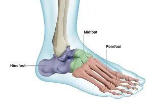
Deconstructing the Bones of Your Foot
While functioning as a single unit, the foot is anatomically divided into three sections: the hindfoot, midfoot, and forefoot.
-
Hindfoot: This section creates the foundation of your ankle and heel. It consists of two primary bones:
- Talus: As mentioned, it’s part of the ankle joint and also extends into the hindfoot.
- Calcaneus: The heel bone, the largest bone in your foot, bearing much of your weight.
-
Midfoot: Acting as a bridge, the midfoot connects the hindfoot to the forefoot. It’s composed of five bones:
- Navicular: Located in front of the talus (ankle bone).
- Cuboid: Situated on the outer side of the foot.
- Cuneiforms (Medial, Intermediate, Lateral): Three wedge-shaped bones arranged in front of the navicular bone on the inner side of the foot.
-
Forefoot: This is the front part of your foot, including the arch and toes. It contains:
- Metatarsals: Five long bones extending from the midfoot to the base of the toes, forming the arch of your foot which is vital for shock absorption during movement.
- Phalanges: These are the bones of your toes (digits). Each toe has three phalanges (proximal, middle, distal) except for the big toe (hallux), which has only two (proximal and distal).
- Sesamoid Bones: Two small, round bones located beneath the big toe joint, embedded in tendons. They assist in the big toe’s movement and reduce pressure.
Joints: The Points of Motion in Your Ankle and Foot
With 33 joints, the foot and ankle are incredibly flexible. These joints can be categorized by their type of movement:
- Hinge Joint (Ankle Joint – Talocrural joint): Primarily allows for dorsiflexion (bending the foot upwards) and plantarflexion (pointing the foot downwards).
- Gliding Joints (Subtalar and Intertarsal joints in the hindfoot): These joints enable side-to-side and subtle rotational movements, crucial for adapting to uneven surfaces.
- Condyloid Joints (Metatarsophalangeal joints in the forefoot and toes): These allow for flexion, extension, abduction (moving away from the midline of the body), and adduction (moving towards the midline of the body) of the toes.
These numerous joints work in concert to provide stability, bear your body weight, and enable a wide range of movements from walking and running to intricate balancing acts, even on varied terrain.
Soft Tissues: The Supporting Structures
Beyond bones and joints, soft tissues are critical for the function of your feet and ankles. These include cartilage, ligaments, muscles, tendons, and bursae.
-
Articular Cartilage: A smooth, resilient tissue lining the joint surfaces of all bones in the foot and ankle. It acts as a shock absorber and minimizes friction between bones, allowing for pain-free movement. Synovial fluid further lubricates this cartilage.
-
Ligaments: Tough, fibrous bands that connect bones to bones. They are crucial for joint stability and preventing excessive movement.
- Plantar Fascia: The largest ligament in the foot, running along the sole from the heel to the forefoot. It supports the arch of the foot and plays a key role in walking and running.
- Lateral Ligaments: Located on the outer ankle, providing stability against excessive inversion (turning the sole inward).
- Medial Ligaments (Deltoid Ligaments): Found on the inner ankle, resisting excessive eversion (turning the sole outward).
-
Muscles: Approximately 20 muscles in the foot contribute to movement and support. Key muscles include:
- Anterior Tibial Muscle: Responsible for dorsiflexion (lifting the foot up).
- Posterior Tibial Muscle: Supports the arch of the foot and assists in plantarflexion.
- Peroneal Muscles (Longus and Brevis): Control eversion and stabilize the ankle on the outer side.
- Extensor Muscles (Extensor Digitorum Longus and Hallucis Longus): Lift the toes up, essential for clearing the ground when walking.
- Flexor Muscles (Flexor Digitorum Longus and Hallucis Longus): Curl the toes downwards and provide stability while standing.
- Intrinsic Foot Muscles: Smaller muscles within the foot that fine-tune toe movements and arch support.
-
Tendons: Fibrous cords that connect muscles to bones, transmitting the force of muscle contraction to create movement.
- Achilles Tendon: The strongest tendon in the body, connecting the calf muscles to the heel bone (calcaneus). It is crucial for plantarflexion, especially during push-off activities.
- Peroneal Tendons: Run along the outer ankle, supporting eversion.
- Tibialis Anterior and Posterior Tendons: Run along the front and back of the ankle respectively, contributing to dorsiflexion and plantarflexion.
-
Bursae: Small, fluid-filled sacs located between tendons and bones or skin. They reduce friction and allow for smooth movement of tendons over bony prominences.
Understanding the intricate anatomy of your feet and ankles highlights their complexity and vulnerability. If you’re experiencing pain, discomfort, or limited mobility, seeking expert care is crucial. Searching for a “foot and ankle doctor near me” is the first step towards addressing your concerns and maintaining the health of these essential structures for an active and pain-free life.